
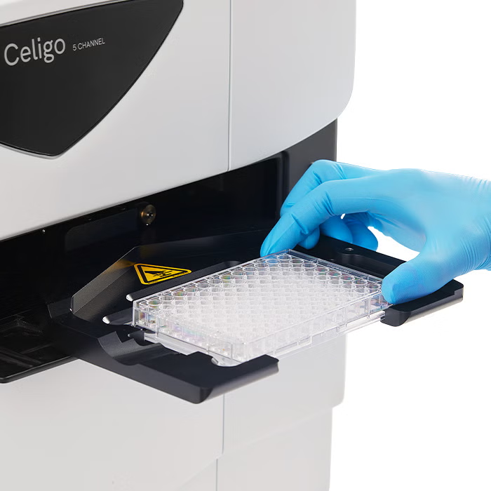
Performs direct cell quantification without trypsinization for adherent cells or cell removal, maintaining experimental conditions.
Supports microplates from 6-well to 1536-well formats as well as T-flask and petri dish compatibility for maximum experimental flexibility.
Captures complete well images with proprietary optics providing uniform illumination and excellent edge-to-edge contrast for cells in plate.
Images and analyzes entire 384-well plates in less than 2 minutes with minimal plate movement, ensuring sample integrity.
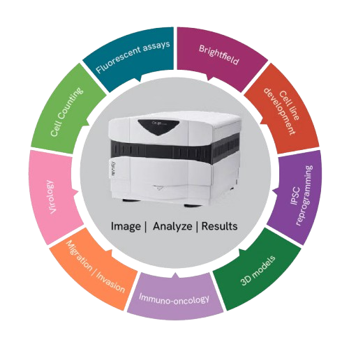
Features brightfield plus 4 fluorescent channels (Blue, Green, Red, Far-Red) for comprehensive cell analysis and multi-parameter assays.
Easily integrates with robotic systems, plate stackers, and liquid handlers for high-throughput screening workflows.
Specialized capabilities for spheroid growth tracking, viability assessment, and drug screening in physiologically relevant 3D cultures.
Performs cell-mediated cytotoxicity assays (ADCC, NK cell killing, CAR-T) with individual cell-level sensitivity.
Provides single cell detection, single cell to single colony verification, and transfection/transduction efficiency monitoring.
Performs time-lapse studies for growth tracking, migration assays, and real-time monitoring of cellular processes.
Celigo is a plate-based benchtop brightfield and fluorescent imaging system designed for whole-well live-cell analysis and cell sample characterization. The Celigo system images and analyzes cells in various types of vessels including 6 – 1536 well plates, T25, T75 flasks, 10 cm dishes, and glass slides without disturbing their natural state.
Individual cell level analysis is easily generated, providing cell level insights unlike ELISA or protein-based assays, and at a faster rate than flow cytometry. A broad range of complex cell-based assays have been optimized for the Celigo cytometer.
Excellent optics for enhanced image quality. Improves brightfield optical image quality at the edge of wells and reduces edge optical distortion by using an F-Theta lens for superior well edge-to-edge image contrast.
Analyze your cell sample without trypsinization to help avoid losing cells and look at cells right where they grow over multiple scan times.