

Brochure
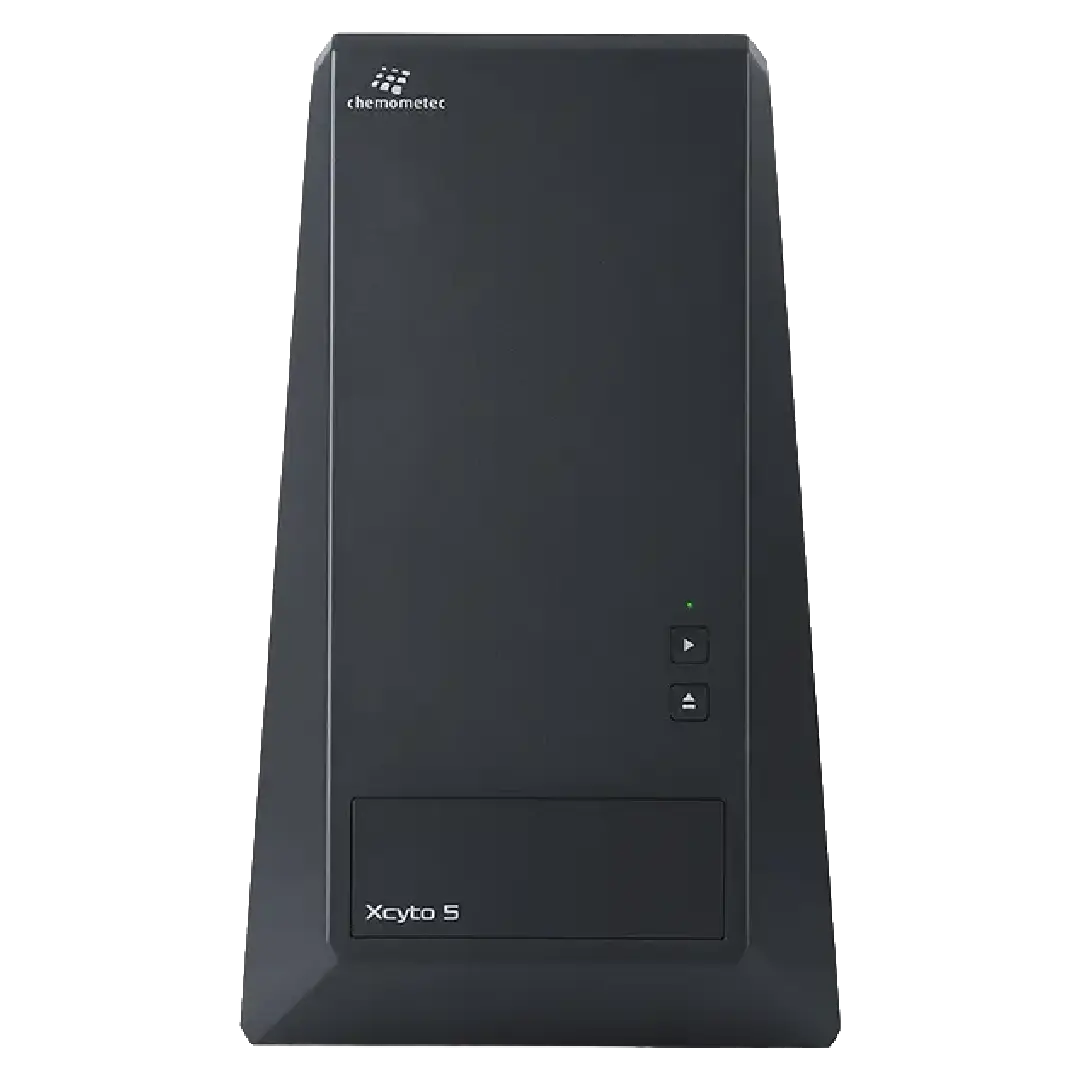
Step into the future of cell analysis with the Xcyto® 5, a compact and user-friendly image cytometer that combines simplicity and precision. Whether you’re in R&D, clinical research, manufacturing, or quality control, the Xcyto® 5 has you covered with its advanced brightfield imaging and wide range of fluorophores.
Imagine effortlessly analyzing both adherent and suspension human and animal cells, as well as yeasts, without the hassle of warm-up times or clogging issues. The Xcyto® 5 delivers flow-cytometry-like data, making your workflow smoother and more efficient.
With its user-friendly and AI-powered software, you can easily set up protocols, view, visualize, and analyze your samples. The Xcyto® 5 is 21 CFR Part 11/GMP ready, seamlessly integrating into your clinical workflow. Just load your cells, start the analysis, and watch as your lab experience is elevated to new heights.
Revolutionize your lab experience with the Xcyto® 5—where innovation meets simplicity.
| System description | Highly sensitivity multicolor image cytometer for suspension and adherent cells. Fully automated production of qualitative as well as quantitative data of individual cells. |
| Camera | CMOS 5.0 MP camera (2448 x 2048 pixels), pixel size: 3.45 µm |
| Light sources | 4 LEDs (405, 488, 535 and 635 nm) for fluorescence and scatter imaging, 1 UV LED (365 nm) for transmitted light microscopy |
| Imaging channels | 25 fluorescent channels employing 9 emission filters (standard configuration), 5 non-fluorescent channels |
| Objective | 4x (NA: 0.13) |
| Image resolution | 0.9 µm/pixel |
| Focus | Contrast-based autofocus |
| Stage | Automated X-Y scanning stage |
| Sample formats | Xcyto® slides and culture slides |
| Cell and culture types | Most adherent and suspended human, animal and yeast cells |
| Data acquisition and analysis | Fully automated image capture integrated with image analysis, comprising cell segmentation, feature extraction and data plotting |
| Analysis time | 20,000 cells/min/channel at 5E6 cells/ml |
| File output | .tif, .png, .jpg, .bmp, .fcs and .acs |
| Detection limit | <2000 FITC molecules |
| Fluorescence resolution (precision) | Coefficient of variation < 3% |
| Dynamic range | > 4 decades |
| Fluorescence linearity | CF405S, CF488A, CF568 and CF647 R2 > 0.995. 4n/2n ratio of 1.95-2.05 for DAPI and PI stained CEN |
| Dimensions and weight | 300 x 500 x 410 mm (W x H x D), 25 kg |
| Input voltage/power consumption | 100-240 V (50-60 Hz)/150 W |
| Operation and storage conditions | Ambient temperature 15°C – 35°C, Relative humidity 10% to 80%, noncondensing |
| Product type | Product No. | Product name |
| Sampling devices | 942-0004 | Xcyto® 2-Sample Slide |
| 942-0010 | Xcyto® 2-Chamber Slide | |
| Software | 950-5001 | XcytoView™ |
| Solutions | 910-3012 | Solution 12 |
| 910-3019 | Solution 19 | |
| 910-3021 | Solution 21 | |
| Accessories | 930-0006 | Power supply |
| 931-0061 | USb 3.0 cable (Type A to MicroB, 3m) |