Read MoreReal-Time Imaging and Automated Quantification of Wound Closure Dynamics with Celloger® ProArticleCelloger® Pro enables automated, real-time analysis of wound healing assays with high accuracy and reproducibility. It simplifies cell migration studies and improves consistency in results.Integrated Solution For Optimizing Lab AutomationCuriosis provide innovative lab automation solutions that maximize accuracy and productivity, enabling faster and more precise outcomes.Contact Usarrow_forwardarrow_back
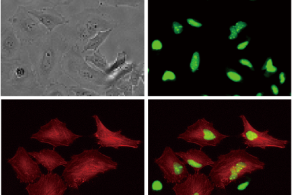
Multicolor fluorescence and brightfield imaging
With its dual color fluorescence and bright-field imaging capabilities, Celloger® Pro enables the capture of high-quality and high-resolution images.
With enhanced scanning methods and innovative merging techniques, the system reduces scanning time, enabling researchers to analyze cellular dynamics with exceptional clarity and efficiency.
Research Applications
Actin dynamics
HeLa cells expressing tdTomato-actin were treated with 1.25 μM and 10 μM cytochalasin B, with 1 hour interval images taken over 48 hours.
Adipogenesis
Observe adipocyte differentiation in 3T3-L1 cells stained with LipiDye II over 48 hours, capturing 1-hour interval images.
NK cell killing assay
The U-2OS cells(target) were stained with CellTracker Green CMFDA and then co-cultured with NK-92 cells to observe the process of NK cells killing the target.
Spheroid cytotoxicity
Monitor the drug effect on HEK293-GFP spheroids treated with Staurosporine by capturing images every 30 minutes for 24 hours.
Transfection efficiency
To monitor gene transfection efficiency in real-time, AGS cells were stained with CMFDA dye and then transfected with the tdTomato-Lifeact gene.
Our Expertise
Automated live cell imaging system
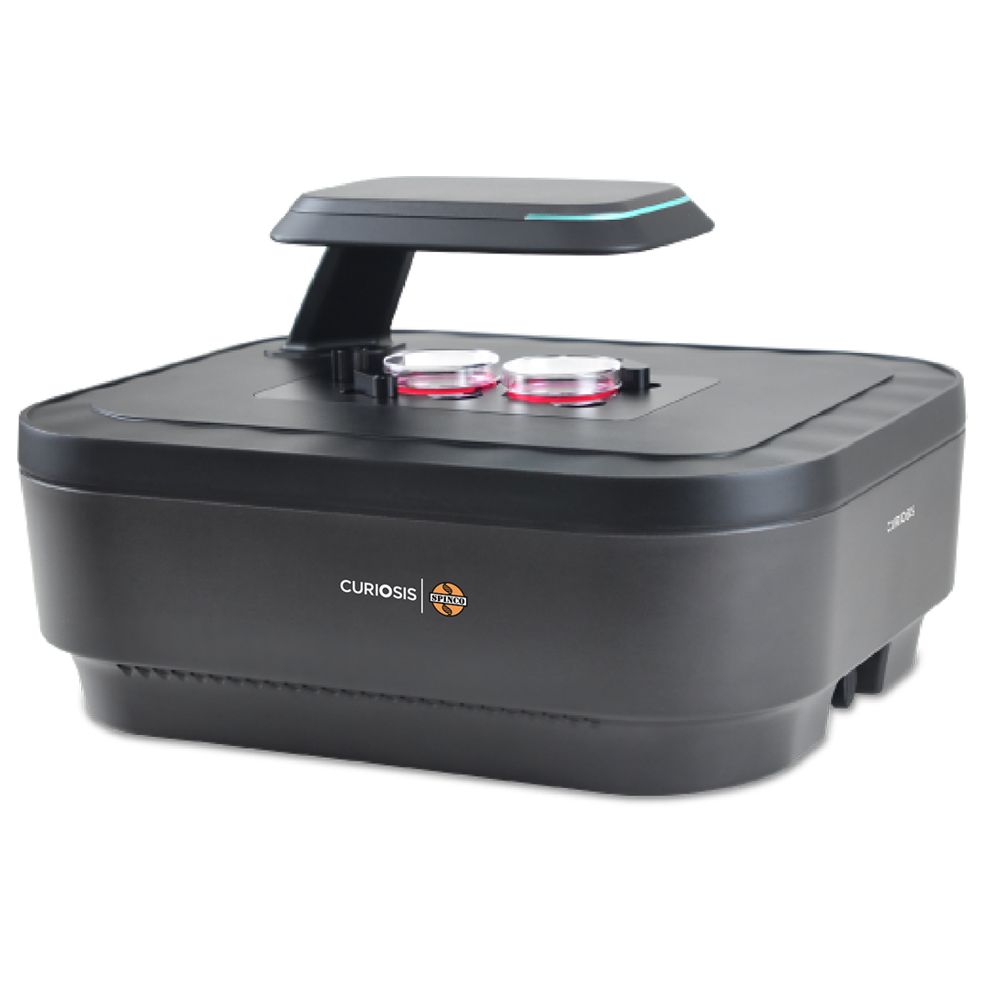
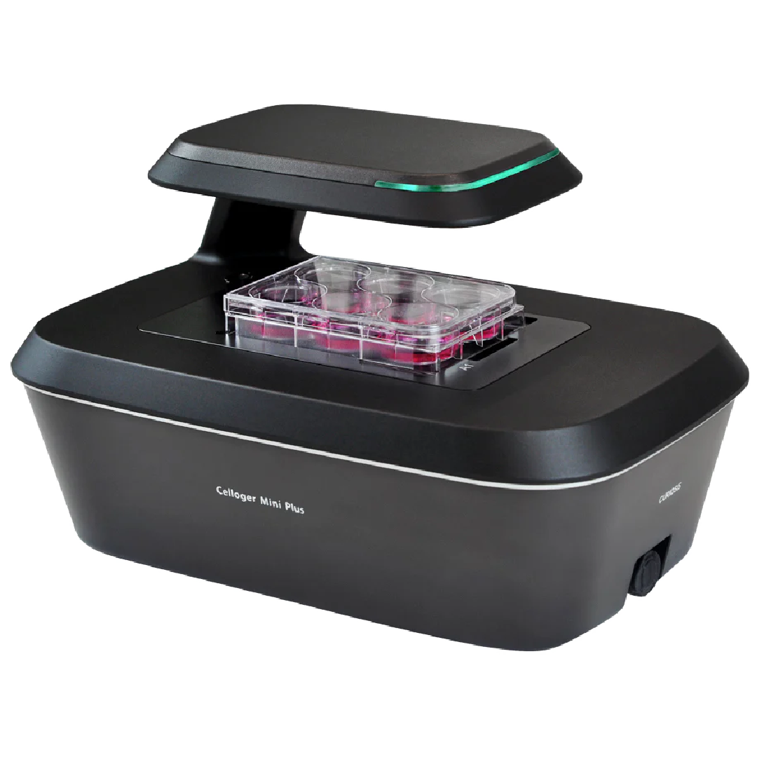
Celloger® Pro is an advanced live cell imaging system offering exceptional image quality and convenience. It enables real-time cell monitoring inside the incubator, allowing seamless observation and tracking of cellular dynamics without disturbing the natural growth environment. With dual fluorescence and bright-field microscopy, multiple markers can be visualized simultaneously, while multi-point time-lapse imaging captures dynamic cellular events at various locations.