Read MoreRapid, quantitative, multi-attribute mass photometry analysis for mRNA stability studiesAPPLICATION NOTEMass photometry enables rapid, high-resolution analysis of mRNA integrity and purity under native conditions, providing single-molecule quantification of transcripts, fragments, and aggregates, detecting stability differences and low-abundance species more accurately than CGE, and streamlining workflows for robust mRNA therapeutic quality assessment.Accurately measure the mass of single biomolecules in solution, in their native state, and without the need for labels.Mass photometryContact Usarrow_forwardarrow_back
Illuminating single biomolecules capturing greater insights
Mass photometry is a simple, easy-to-use technology that rapidly analyzes biomolecules at the single-particle level in solution. It measures the light scattered by individual particles and uses the signal to count the particles and measure their mass with high accuracy.
Mass photometry measurements provide the mass distribution and relative concentrations of a wide range of biomolecules in a sample, accelerating a variety of studies, including:
Sample purity and homogeneity
Protein oligomerization
Biomolecular interactions
Macromolecular complex assembly
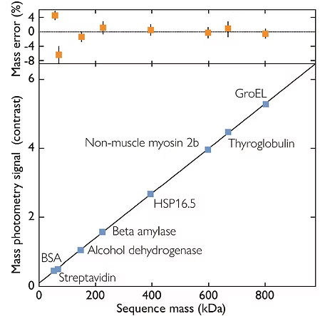
Figure1: The amount of light scattered by a particle scales linearly with the particle’s volume and refractive index. As the optical properties and density of biomolecules vary only by just a few percent, the scattering signal is directly proportional to the molecule’s mass – making it possible to weigh single molecules with light (Figure). The correlation of scattering signal with mass holds true for a variety of biomolecules (e.g. glycoproteins, nucleic acids and / or lipids), making mass photometry a universal analysis tool for biomolecules in solution.
Benefits of Mass Photometry
Efficient bioanalysis of molecules in their native state
Accurate mass measurement of true native behavior
In solution, in a variety of buffers and compatible with membrane proteins
Label-free, without the need to modify samples
Information on all sub-populations in samples
Single-molecule counting
Wide mass range and high dynamic range
One assay format delivering multiple results
Homogeneity, structural integrity and activity
Quick, simple, minimal sample amounts
How does mass photometry work?
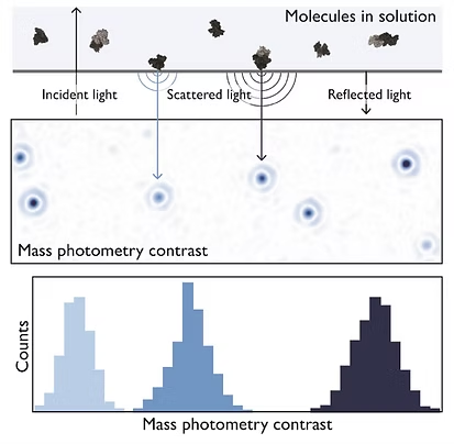
Mass photometry is a new way to analyze biomolecules, built upon the principles of interference reflection microscopy and interferometric scattering microscopy. Mass photometry enables the reliable detection of single molecules and the direct measurement of each molecule’s mass in solution.
With a wide dynamic range, mass photometry can be used to assess:
Proteins / glycoproteins
Nucleic Acids
Adeno-associated viruses (AAVs)
Antibodies
VLPs
Membrane proteins
Figure 2: The principle of mass photometry. The light scattered by a molecule that has landed on a measurement surface interferes with light reflected by that surface. The interference signal scales linearly with mass.
What can you do with mass photometry?
Our technology is transforming research in a wide range of fields
AAVs
Precisely characterize AAV preparations by accurately determining empty versus full capsid ratios, aggregation states, and overall integrity. Crucial for optimizing production and ensuring the safety and efficacy of gene therapies.
AdVs
Gain direct, label-free insights into AdV quality by assessing particle concentration, aggregation, and purity. This enables better control over manufacturing processes for gene delivery vectors and oncolytic agents.
Antibody aggregation
Accurately detect and quantify antibody aggregates in solution – faster and with far less sample than SEC and AUC. Essential for optimizing formulation conditions, improving stability, and ensuring safe, effective biotherapeutics.
Antibody characterization
Comprehensively profile bispecific and multi-specific antibody-antigen interactions, including stoichiometry. Ideal for new modalities, complements SPR and BLI, aiding experimental design and the interpretation of data on multi-component systems.
Membrane protein analytics
Analyze membrane proteins in native-like environments, determining oligomeric states, complex formation, and sample purity. Critical for advancing structural studies, functional assays, and therapeutic development.
mRNA
Characterize mRNA integrity and its formulation in lipid nanoparticles (LNPs). Assess size distribution, encapsulation efficiency, and stability of mRNA-LNP complexes, vital for developing and quality controlling mRNA-based vaccines and therapies.
Protein interactions
Directly measure complex formation, stoichiometry, and binding strength to reveal how proteins interact in solution. Vital for understanding biological mechanisms, validating targets, and accelerating drug discovery.
Lentivirus populations
Our advanced single‑particle analysis reveals population heterogeneity and sample purity. It delivers precise, multiparametric data by simultaneously measuring particle size and mass-related contrast.
our expertise
Clarity. Speed. Simplicity.
At the heart of Refeyn’s innovation lies a commitment to providing truly insightful biomolecular data without the traditional trade-offs. Our mass photometry platform is engineered to deliver clear, actionable results quickly and simply, fundamentally changing how you approach characterization.
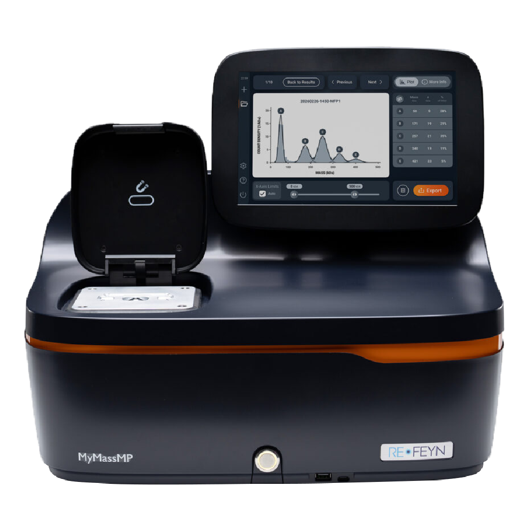
Our new MyMass mass photometer delivers fast, accurate assessment of purity and homogeneity so you can move forward with cryo-EM, native MS, SPR or BLI with confidence.