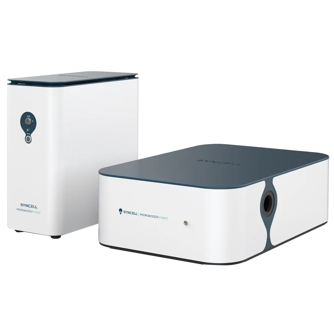
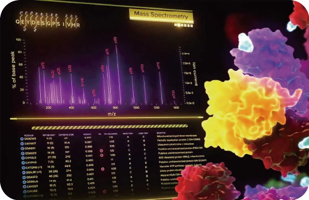
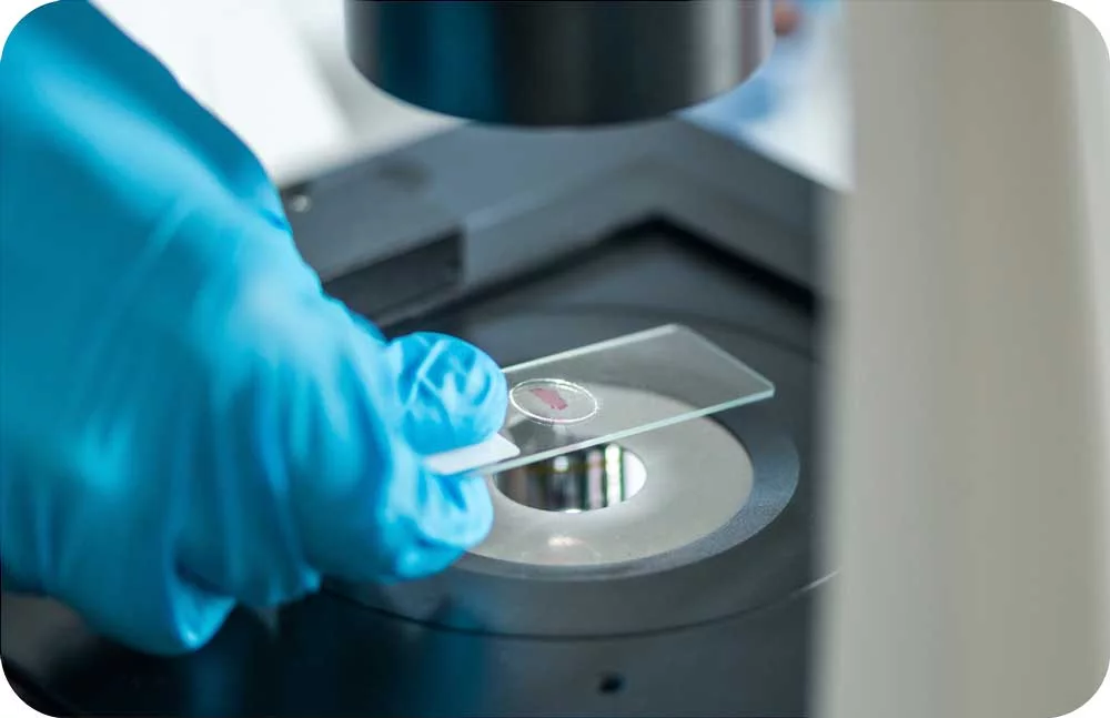
Microscoop® Mint uses microscopy-guided photo-biotinylation to enable superior resolution, specificity, sensitivity, and efficiency.
A uniquely advanced solution to prep for mass spec analysis, the platform’s capabilities and streamlined workflows offers advantages over traditional methods like laser capture microdissection (LCM), antibody-based imaging workflows, and proximity labeling.
Highlights
Explore cellular and subcellular interactions that shape health and disease like never before.
Maximize the value and impact of your mass spec data
Traditional proteomic technology often relies on predefined panels or antibody-based enrichment, which introduces bias—only known or expected proteins are detected.
But Syncell’s Microscoop® labels and analyzes all proteins in a specific region of interest without pre-selection, allowing mass spec to capture low-abundance or rare proteoforms, and reveal otherwise unknown protein constituents with higher dynamic range and specificity than laser microdissection or proximity labeling methods.
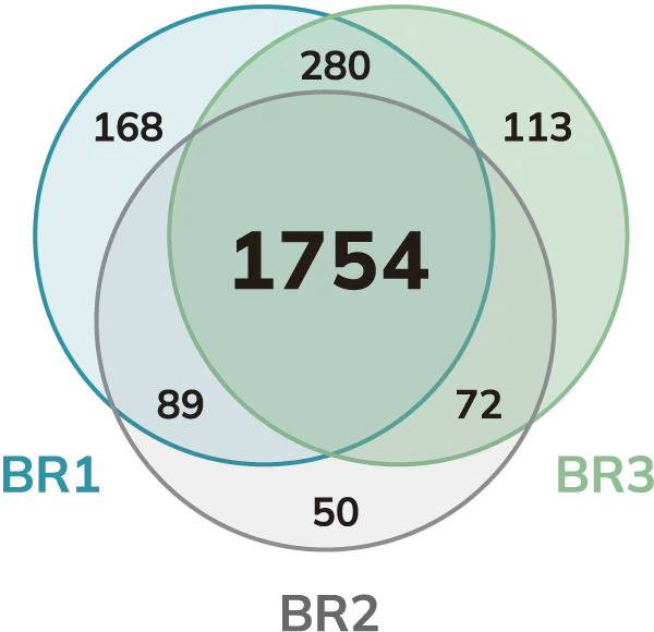
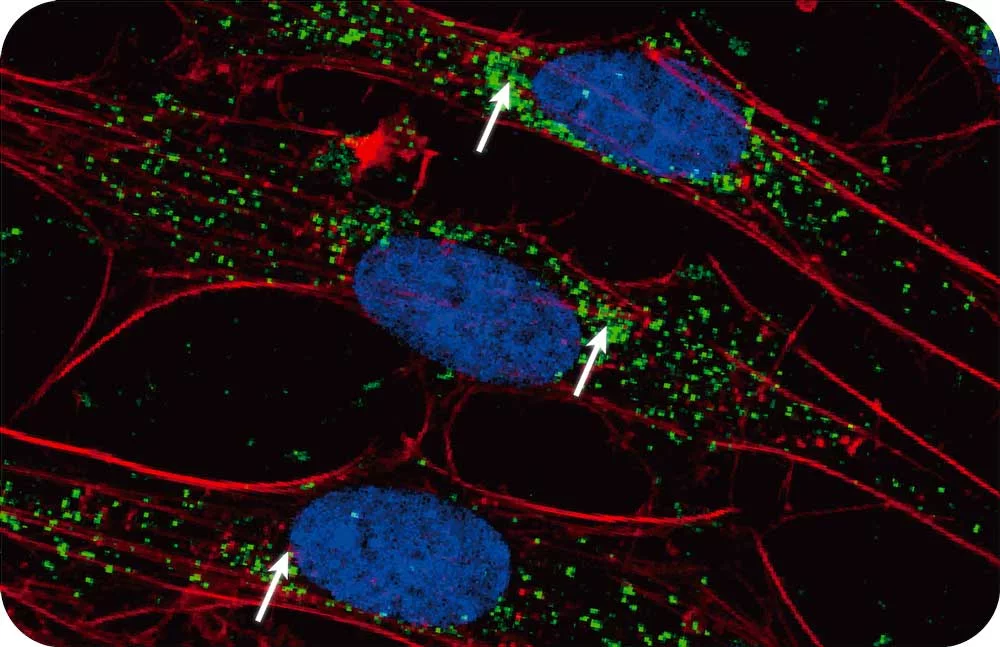
Venn diagram of the stress granule proteins spatially isolated by Microscoop® and analyzed by mass spectrometry.
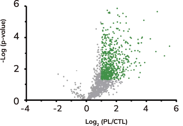
Volcano plot of protein levels in relative photolabeled (PL) samples to control (CTL) samples in log2 scale. Over-represented (enriched) proteins are shown in green.
Spatial Context
Mass spec alone doesn’t provide spatial information but combining it with the power of Syncell’s microscopy-guided photo-biotinylation technology enables researchers to link spatial location with the entire local proteome allowing you to:
understand the entire proteomic makeup of key organelles to identify novel biomarkers (e.g., cilia, nuclei, tumor microenvironments).
understand the location-function relationships of proteins.
identify disease-related signatures based on tissue architecture to hone in on the exact proteomic targets that are responsible for disease progression
High Quality Mass Spec Data
By restricting the proteomic analysis to defined cellular or subcellular regions, our proprietary opto-proteomic platform improves:
signal-to-noise in complex tissue samples
dynamic range and loses low expressing proteins in LC/MS data
and enables single-region proteomic profiling
Sample Access
Microscoop can work with FFPE/fresh frozen tissue, preserving the integrity of archived clinical samples. This brings MS-based insights into areas where methods like proximity labeling are not capable: