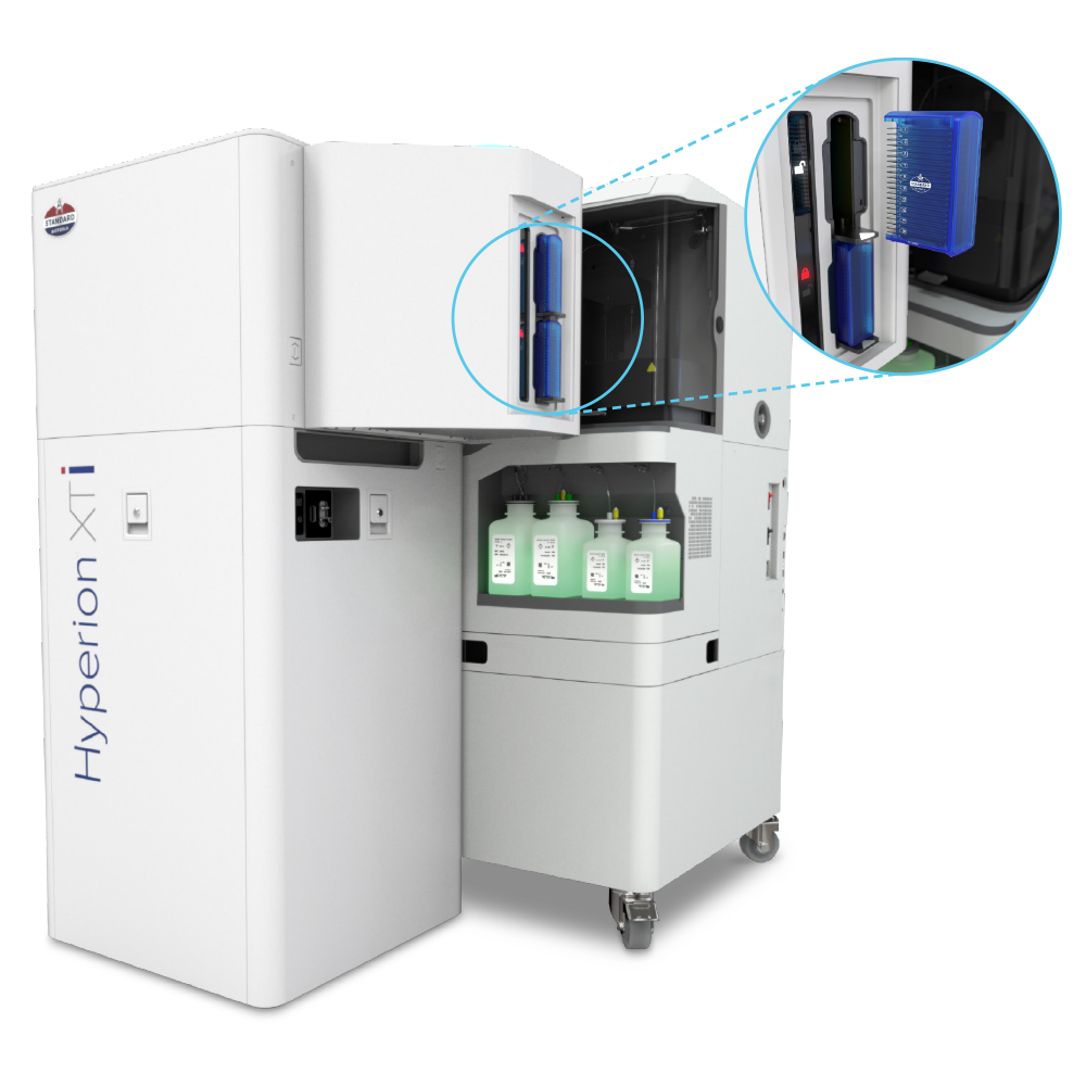
High-throughput multiplexed whole slide tissue imaging: The Hyperion XTi Imaging System now offers the ability to automate slide loading for continuous acquisition and new whole slide imaging modes for rapid and detailed analysis.
Leverage the unique integrated dual modality of single-cell and spatial proteomic capabilities with the Hyperion XTi Imaging System and CyTOF XT systems. Take a deep dive into translational and clinical applications that explore new views of cellular composition within the tissue microenvironment. Watch this brief overview.
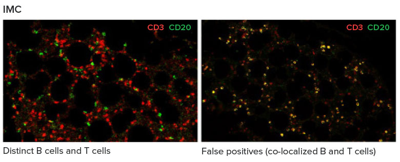
Powered by proven Imaging Mass Cytometry™ (IMC™) technology, the Hyperion XTi Imaging System is a multiplexed tissue imager that uses metal-tagged antibodies, instead of fluorophores, to simultaneously and reliably acquire 40-plus protein and RNA markers, without autofluorescence interference. IMC technology is the trusted choice for researchers and continues to be featured in high-impact publications – making it the most reliable tool for highly multiplexed imaging.
Researchers can visualize 40-plus markers in real time, without managing time-consuming acquisition cycles – expediting the time to deeper analysis.
View the Hyperion XTi image lookbook to learn how whole slide and cell imaging modes can be applied.
Represents Hyperion XTi Preview Mode for a large tissue section over 25 minutes. Researchers can select and view from any markers on the panel during acquisition.
Incorporate a new level of throughput and efficiency with the Hyperion XTi Slide Loader. The optional slide loader enables automatic loading and acquisition of up to 40 slides. It is equipped with a barcoded slide reader for easy sample identification, providing researchers with peace of mind when managing precious tissue samples.