Read MoreUnraveling Metabolic Reprogramming in Cancer with Spatial ProteomicsApplication NoteModular Imaging Mass Cytometry panels identify cell metabolic states across the tumor microenvironmentImaging Mass Cytometry™ (IMC™) is the most trusted technology that enables researchers to accurately assess complex phenotypes and immune spatial interactions in the tissue microenvironment.Spatial ProteomicsContact Usarrow_forwardarrow_back
Imaging Mass Cytometry
Imaging Mass Cytometry technology generates high-dimensional spatial data at subcellular resolution. IMC technology uses cytometry by time-of-flight (on which CyTOF™ systems are based) to overcome the multiplexing limitations of traditional immunohistochemistry (IHC) and immunofluorescence. By applying metal-tagged antibodies instead of fluorochromes, IMC technology has the unique capability to simultaneously stain, acquire and analyze 40-plus markers of interest on a tissue section without interference from autofluorescent tissues or management of spectral overlap.
IMC is the only technology with
No autofluorescence interference to image any tissue type
40-plus markers imaged simultaneously to get results faster
Integrated cell segmentation for faster interpretation
Batch staining of all slides for high-volume studies
Dual imaging and flow cytometry mode to maximize investment
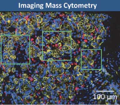
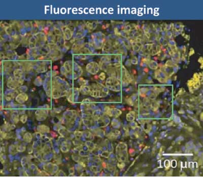
IMC shows the true biology
High-plex imaging for all tissue types ― including lung, bone marrow, colon and brain ― without autofluorescence interference.
Well-defined red signals from CD68
Cellular structure is sharply defined by yellow pan-cytokeratin stain
CD68 indistinct or missing
Cellular structure diffuse
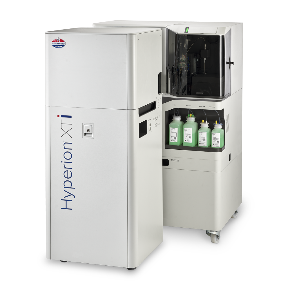
Hyperion XTi Imaging System
Powered by proven Imaging Mass Cytometry™ (IMC™) technology, the Hyperion XTi Imaging System is a multiplexed tissue imager that uses metal-tagged antibodies, instead of fluorophores, to simultaneously and reliably acquire 40-plus protein and RNA markers, without autofluorescence interference. IMC technology is the trusted choice for researchers and continues to be featured in high-impact publications – making it the most reliable tool for highly multiplexed imaging.
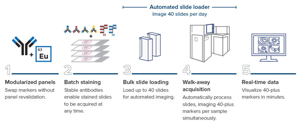
Multiplexed imaging – without compromising speed
An end-to-end workflow to get results faster
CyTOF™ Technology
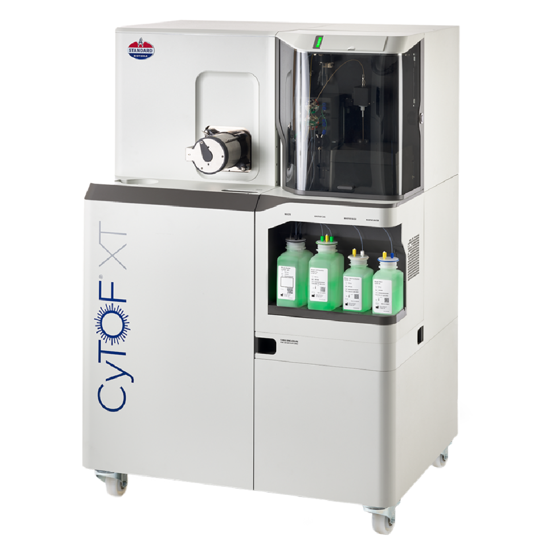
Cytometry by time-of-flight or mass cytometry (on which CyTOF™ technology is based) is a cytometric technique that measures metal-conjugated antibodies bound to cell antigens. It is a pioneering technology developed to overcome many of the challenges experienced with fluorescence flow cytometry, increasing its utility by enabling the simultaneous assessment of a much higher number of parameters. This added capability maximizes the information obtained from each sample and, subsequently, each cell.
Why Mass Cytometry
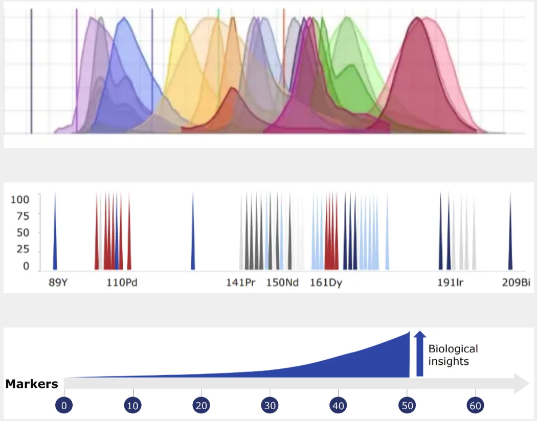
Mass cytometry is advantageous because of its ability to precisely and accurately distinguish between reporter molecules. By using atomic mass measurements, mass cytometry avoids autofluorescence signals from cells and tissues, enables fast panel design since all antibodies are used at once and facilitates barcoding to multiplex samples in large-scale acquisitions.
Mass spectrometry detects many different metal isotopes with high accuracy. By combining the single-cell analysis approach of flow cytometry with the precision of mass spectrometry, mass cytometry enables high-parameter analysis while generating little to no signal overlap. This eliminates the need to correct fluorescence spillover and further enables the use of statistical techniques to facilitate screening drug candidates and assessing on- or off-target effects post-treatment.
Find the solution that fits your research
Panel designer
Build an optimized panel
Maxpar™ Direct Immune Profiling Assay
Standardized immune profiling in a single tube
Quick-start imaging panels
Ready-to-use Imaging Mass Cytometry™ panels
Maxpar Panels
Easy immune profiling with pre-validated panels
End-to-end solutions
Lab Services support for any project
Custom genomic assays
D3™ Assay Design with expert support
Lyophilized CyTOF panels
LyoMax™ Panels for stability and speed in high-volume studies
our expertise
Our mission
Unleashing tools to accelerate breakthroughs in human health
Standard BioTools delivers the leading omics solutions that empower life sciences researchers to discover unique biological insights not accessible by other technologies.